- Printed Journal
- Indexed Journal
- Refereed Journal
- Peer Reviewed Journal
P-ISSN: 2349-6800, E-ISSN: 2320-7078


Journal of Entomology and Zoology Studies
2016, Vol. 4, Issue 3
The histological relationship between the environmental stress and milk production in Iraqi Buffaloes; Bubalus bubalis
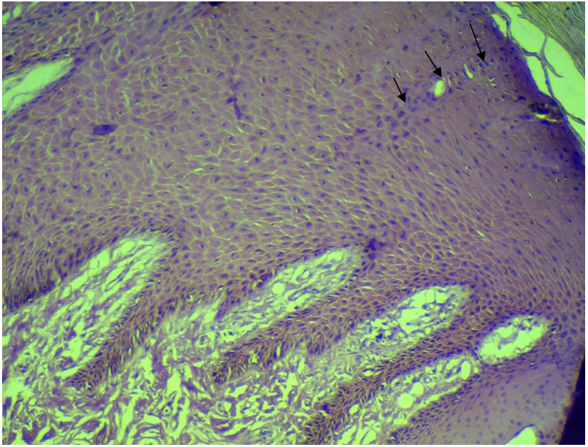
Fig. 1: Epidermis of skin of buffalo. Note the direction of one excretory duct of the sweat gland toward the surface of the skin (arrows). H& E stain.X40.
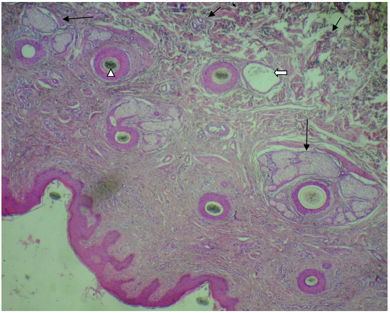
Fig. 2: Skin of buffaloes. Note the small deep acini of sweat glands (short black arrows), their ducts (white arrow), the abundant melanocytes (white arrow head) and the lobulated sebaceous glands (Long black arrow). H & E stain. X 100.
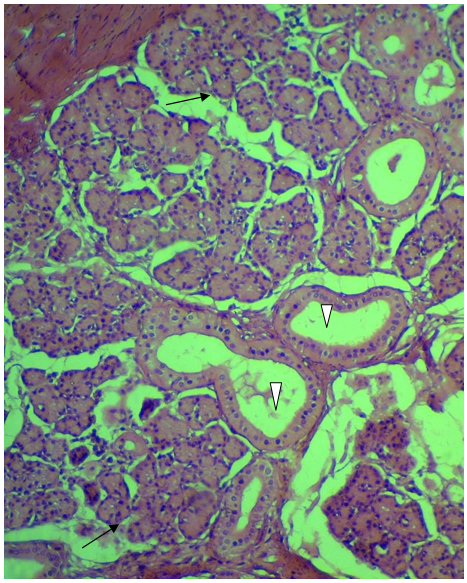
Fig. 3: High magnification of buffaloe skin dermis refers to the secretory acini (black arrows) and their duct system (white arrow heads) of sweat glands. H&E stain. X 400.
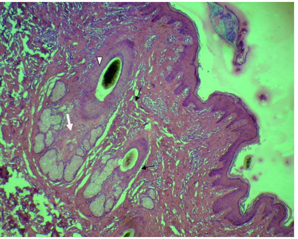
Fig. 4: Skin of buffaloe. Note the distribution of simple sweat glands (black arrows) and sebaceous glands (white arrow).The dark spot (white arrow head) refers to the melanocyte within the hair follicle. H & E stain. x200.
Indexed In

Hard Copy Subscription
Click Here for more InformationOur Other Journal

Important Topics
Related Links
Related Journal Subscription
Important Publications Links
Important Links

Journal of Entomology and Zoology Studies

- Home
- Editorial Board
- Archives
- Instructions
- Membership
- Publication Ethics
- Publish Book (ISBN)
- Make Payment
- Contact Us
- Helpline No.: +91-9711224068
- Fast Publication: +91-7048922346
- Toll Free: 1800-1234070
