Advertisement:
Journal of Entomology and Zoology Studies
Volume 1, Issue 4
Determining with SEM, structure of the venom apparatus in the tube web spider, Segestria florentina
(Araneae: Segestriidae)
Author(s): Mehlika Benli 1*, Nazife Yigit1, Mehmet Karakas1 and Suna Cebesoy1
1. Ankara University, Faculty of Science, Department of Biology, 06100 Tandogan, Ankara– TURKEY
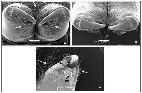
Abstract: The objective of the present study was to describe functional morphological features of venom apparatus in the tube web spider, Segestria florentina (Rossi, 1790) (Araneae: Segestriidae) by using scanning electron microscope (SEM). The venom apparatus is situated in the anterior part of the prosoma, and is composed of a pair of venom glands and chelicerae. The chelicera of S. florentina has two parts: basal segment and a movable articulated apical segment (fang). The cheliceral fang rests in a groove on the basal segment of chelicerae. A venom hole is located on the subterminal part of each fang. A pair of venom glands is completely separate but similar to each other within the prosoma.Each venom gland is surrounded by striated muscle bundles, such as with the capsules. The venom, produced in the venom glands, is carried by venom ducts passing throughout the chelicerae. Each venom gland has its own venom duct, chelicera and fang. The venom is excreted from the venom pore on the subterminal part of the fang by means of muscular contractions covering the venom gland.
Download Full Article : Click Here
Advertisement: