- Printed Journal
- Indexed Journal
- Refereed Journal
- Peer Reviewed Journal
P-ISSN: 2349-6800, E-ISSN: 2320-7078


Journal of Entomology and Zoology Studies
2013, Vol. 1, Issue 4
Morphology and Histology of the Male Reproductive System in Graphosoma lineatum (Heteroptera: Pentatomidae) Based on Optical and Scanning Electron Microscopy
Nurcan Özyurt 1, Selami Candan 1, Zekiye Suludere 1, Damla Amutkan 1
The male reproductive system of Graphosoma lineatum (Linnaeus 1758) is studied morphologically and histologically using both light and scanning electron microscopes (SEM). On the basis of histological sections and whole morfological studies, the following elements in the structure of the male reproductive system of G. lineatum have been examined: the structure of a pair of testis containing the testes follicles and the stage of spermatogenesis, the connection of testes and vas deferens; the position and the histological structure the of a pair of vas deferens which is the tube down which the sperm travels, the structure of seminal vesicle which is where the sperm is stored prior to mating which supply seminal fluid for additional volume and to nourish the sperm before-during their journey, the bulbus ejaculatorius, a pair of ectodermal sacs the ductus ejaculatorius as well as the structure of aedeagus.
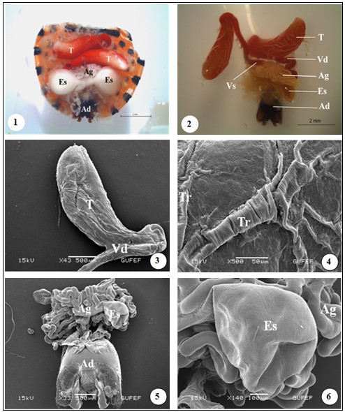
Fig. 1: Light photos of general view of the male reproductive system of G. lineatum. Testes (T), vas deferens (Vd), seminal vesicles (Vs), accessory glands (Ag), ectodermal sac (Es), aedeagus (Ad). Figs 3-4: SEM photos of vas deferens and trachea (Tr) on the surface of the testes. Fig 5: SEM picture of accessory glands, ectodermal sacs, aedeagus. Fig 6: SEM photo of ectodermal sac and accessory glands
Pages : 40-46 | 2386 Views | 306 Downloads


How to cite this article:
Nurcan Özyurt 1, Selami Candan 1, Zekiye Suludere 1, Damla Amutkan 1. Morphology and Histology of the Male Reproductive System in Graphosoma lineatum (Heteroptera: Pentatomidae) Based on Optical and Scanning Electron Microscopy. J Entomol Zool Stud 2013;1(4):40-46.
Indexed In

Hard Copy Subscription
Click Here for more InformationOur Other Journal

Important Topics
Related Links
Related Journal Subscription
Important Publications Links
Important Links

Journal of Entomology and Zoology Studies

- Home
- Editorial Board
- Archives
- Instructions
- Membership
- Publication Ethics
- Publish Book (ISBN)
- Make Payment
- Contact Us
- Helpline No.: +91-9711224068
- Fast Publication: +91-7048922346
- Toll Free: 1800-1234070
