- Printed Journal
- Indexed Journal
- Refereed Journal
- Peer Reviewed Journal
P-ISSN: 2349-6800, E-ISSN: 2320-7078


Journal of Entomology and Zoology Studies
2019, Vol. 7, Issue 6
Pathological observation of incidental Spirocerca lupi infection with associated spontaneous cellulitis in a mongrel dog
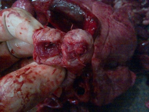
Fig. 1: Cut out tumorous nodule containing Spirocerca lupi parasites (arrow)
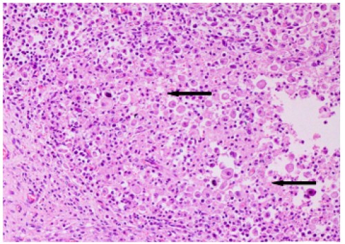
Fig. 2: Esophagus, wall showing parasitic eggs surrounded by degenerated and viable swollen macrophages and eosinophils, H&E x20.
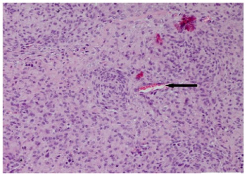
Fig. 3: Esophagus, wall showing diffuse presence of fibroblast in “whorl” pattern all throughout the section (arrows), H&E x 20
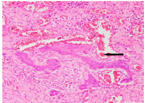
Fig. 4: Esophagus, wall, osseous transformation of fibroblastic cells (arrow). H&E x20.
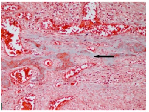
Fig. 5: Esophagus, wall, partial mineralization of osteoid mass (arrow). Masson trichrome x20
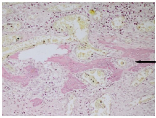
Fig. 6: Esophagus, wall with well demarcated osteoid zone (arrow). Van Geison x 20
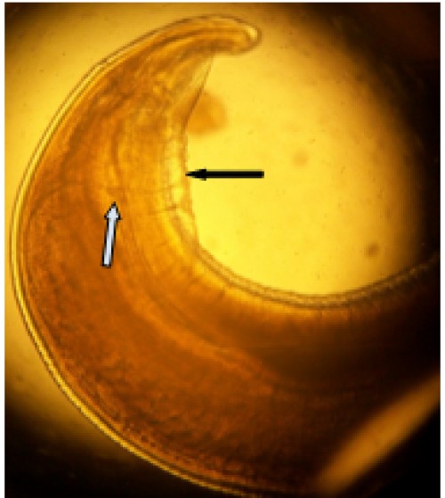
Fig. 7: Spirocerca lupi, male showing two (2) unequal posterior spicules (white arrow) and four (4) lateral alae
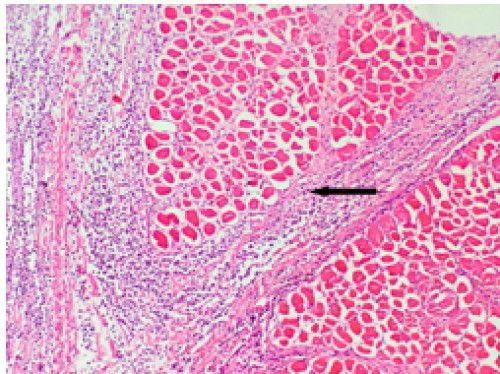
Fig. 8: Subcutaneous tissue and intermuscular fascicles containing purulent exudates, necrotic areas with gradual constriction of overall muscle mass (arrow), H&E x20.
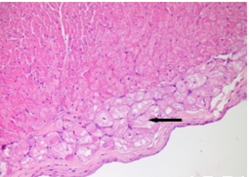
Fig. 9: Heart, vacuolar andgranular degeneration of purkinjee cells, H&E x20
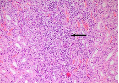
Fig. 10: Kidney, distinct infiltration of lymphocytes, macrophages and lymphoblast in interstitium, H&E x20
Indexed In

Hard Copy Subscription
Click Here for more InformationOur Other Journal

Important Topics
Related Links
Related Journal Subscription
Important Publications Links
Important Links

Journal of Entomology and Zoology Studies

- Home
- Editorial Board
- Archives
- Instructions
- Membership
- Publication Ethics
- Publish Book (ISBN)
- Make Payment
- Contact Us
- Helpline No.: +91-9711224068
- Fast Publication: +91-7048922346
- Toll Free: 1800-1234070
